Cemented total hip prosthesis
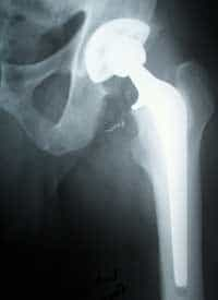
Over the years, but not only, the joints lose elasticity and arthrosis phenomena can occur which cause pain, up to a poor mobility of the patient. In these cases, a hip prosthesis similar to the one visible in the radiograph to the side is used. The procedure itself is quite simple and takes about an hour. The most delicate and complicated phase is the planning of the intervention itself, in which the correct position and inclination of the prosthesis which will replace the bone must be calculated. Planning is done based on x-rays on which transparencies are superimposed with the various prostheses available to find the most suitable size. Misjudgment at this stage could result in incorrect prosthesis angulation, which would result in the patient walking abnormally upon discharge. For the total replacement of the hip joint, non -cemented titanium alloy prostheses are used, which allow the patient to walk again after a maximum of 3 or 4 days after the operation. Furthermore, the non-cementification of the plant is an advantage because the polymer glues used are highly toxic and cause necrosis. [ Continuing in the article, all the main phases of the intervention are illustrated and by their nature they are not suitable for "sensitive" people ]
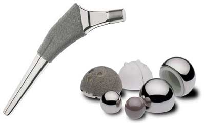
Over a titanium alloy prosthesis. Note the " rough " part of the stem which promotes the osseointegration of the implant. At this point, the data, previously obtained in the preoperative phase, are reported with a felt-tip pen on the patient's leg.
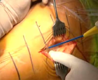
After that we move on to the actual operation by incision of the skin and making the femur come out of its seat.
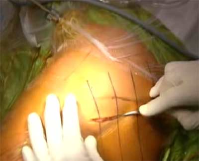
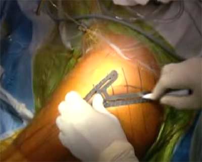
The surgeon uses the electric scalpel and assisted with forceps to approach the femur.
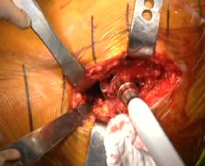
Once the area of interest has been reached, fixed retractors are positioned which guarantee the orthopedic surgeon a wide and stable operating field.
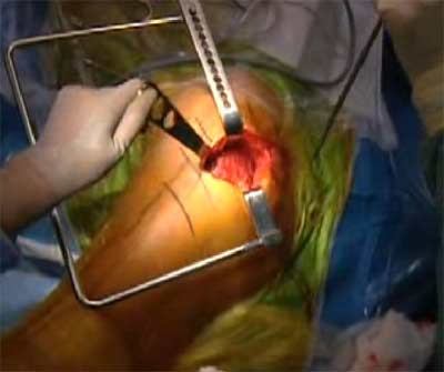
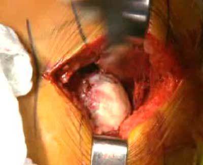
Once the femur is reached, the head is exposed and prepared for the next step.
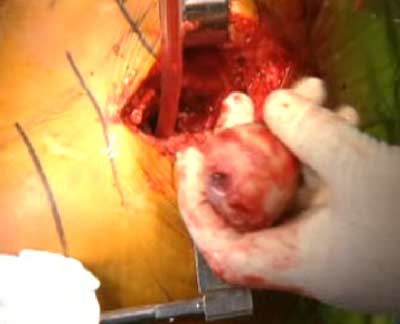
At this point the end of the femur is cut with an electric jigsaw. In the photo the surgeon is holding the head of the femur that has just been removed in his hand.
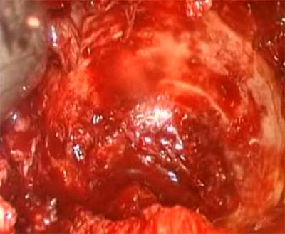
Once the femoral head has been cut, the housing of the acetabular cup is prepared with a kind of drill which scrapes off the cartilage and a part of the bone.
This is the result that is obtained once the scraping is completed.
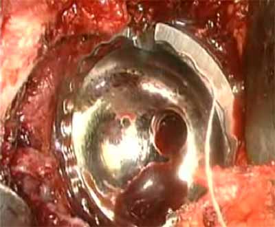
With a mallet, the surgeon fits the acetabular cup into the bone, always checking the correct positioning.
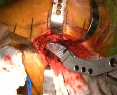
Now the orthopedist drills the femur, thus extracting the marrow, to prepare the femoral canal which will house the stem of the prosthesis. The hole is made smaller than the actual size of the prosthesis, to ensure immediate sealing and cohesion.
Once this operation has been completed, the pressure prosthesis is inserted and the limb is attempted to move to make sure that everything has been done in a workmanlike manner.

Barring unforeseen circumstances, the tissues are repositioned and the cut is sutured. Research at the moment is focused on finding increasingly biocompatible and long-lasting materials. In fact, one of the major limitations is the duration of this device which is currently around 20 years and sometimes even 30. After this period of time, the prosthesis must be re-interventioned and replaced. The second operation is always more difficult than the first, because the bone loses consistency and weakens.
You may also like

MOC, Computerized Bone Mineralometry for osteoporosis
X-ray computerized bone mineralometry (abbreviated as MOC ) represents the current method of reference in the diagnosis of osteoporosis , successfully used at the polyclinic of Milan and in other Italian hospitals. MOC is based on the principle of photonic absorptiometry: a thin beam of X-rays passing through the tissues is absorbed in proportion to… Continua a leggere MOC, Computerized Bone Mineralometry for osteoporosis
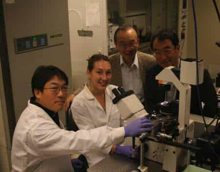
Nanotubes and stem cells for bone reconstruction
It has been found that the use of implants made of titanium nanotubes together with stem cells can accelerate the growth of bone tissue. The group of bioengineers from the University of California – San Diego used nano-biotechnology technologies that made it possible to carry out this experiment. In fact, they were able to place… Continua a leggere Nanotubes and stem cells for bone reconstruction
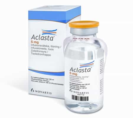
Aclasta against osteoporosis and fractures
Novartis publishes the results of the first study performed on Aclasta , an osteoporosis drug in patients with hip fractures. It has been seen that administering Aclasta once a year prevents new fractures by 35% and improves life in a certain sense. In fact, it would seem that in the group of patients analyzed the… Continua a leggere Aclasta against osteoporosis and fractures

New bone conduction hearing aid
To the aid of those suffering from mixed or conductive hearing loss as well as single sided deafness – Single Sided Deafness (SSD) comes the new Baha system. This brand new hearing prosthetic aid differs from the others currently on the market because, instead of using sound amplification through normal air conduction, it directly stimulates… Continua a leggere New bone conduction hearing aid
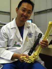
Non-metallic total ankle prosthesis
A study successfully completed and published in the Journal of Foot & Ankle Surgery describing a new technique for ankle repair. the technique developed by Daniel K. Lee, director of foot and ankle surgery at UCSD Medical Center, consists in applying a material very similar to collagen instead of the current titanium or steel prostheses.… Continua a leggere Non-metallic total ankle prosthesis
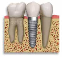
Dental implants covered with synthetic bone
Some researchers will have the opportunity to publish a study on dental coatings in the next issue of the International Journal of Nanomanufacturing. Titanium is the most used material in many dental implants, however its surface is typically inert: this makes it biocompatible and favored to avoid adverse reactions of the immune system, but it… Continua a leggere Dental implants covered with synthetic bone