MOC, Computerized Bone Mineralometry for osteoporosis

X-ray computerized bone mineralometry (abbreviated as MOC ) represents the current method of reference in the diagnosis of osteoporosis , successfully used at the polyclinic of Milan and in other Italian hospitals. MOC is based on the principle of photonic absorptiometry: a thin beam of X-rays passing through the tissues is absorbed in proportion to the density of the components. The subsequent processing of the data, thanks to suitable mathematical algorithms, can calculate the exact density of these tissues, distinguishing between soft and hard tissues (bones). The MOC measures the mineral density of the skeleton, mainly made up of calcium hydroxyapatite crystals, and allows for the evaluation of small variations in bone mineral density over time. 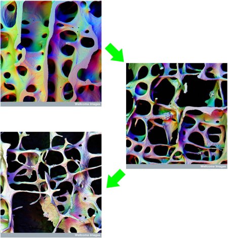 Above three different photos of the section of a lumbar vertebra made with a scanning electron microscope: in the first one above a healthy trabecular bone and then two degrees of osteoporotic degeneration highlighted by the impoverishment of the bone micro-structure. The MOC is a non-invasive exam and does not present risks deriving from radiation for the patient: 10 MOC correspond to the normal daily exposure to background radiation and a flight from Europe to America corresponds to 80 MOC. There are four types of MOC exam:
- Vertebral column;
- Femur;
- Forearm;
- Whole body.
MOC of the spine is particularly indicated in young patients, especially if they have recently undergone menopause, and in short-term monitoring of osteoporosis therapy. The MOC of the femur is instead indicated in elderly patients (over 65 years of age), in which there are frequent calcifications of the abdominal aorta, marked osteoarthritis of the spine, marked dorsolumbar scoliosis, somatic fractures of the lumbar tract, conditions which induce overestimations of quantitative parameters measured with the MOC of the column. The MOC of the spine, the one I will tell you about, is performed with the patient supine on the densitometric table, with a special support under the legs and with the trunk free from all metal objects. The scan is performed on the lumbar spine, takes a few minutes, is painless and does not require specific preparation on the part of the patient. The MOC exam is divided into two parts:
- a first represents the image of the column and is used to check the correct execution of the exam;
- the second presents the quantitative values of bone mineral density, which are the most relevant part of the MOC, among all the total BMD (Bone Mineral Density) value is the most important to consider for the interpretation of the exam.
[pictures wellcome ]
You may also like
Nanotubes and stem cells for bone reconstruction
It has been found that the use of implants made of titanium nanotubes together with stem cells can accelerate the growth of bone tissue. The group of bioengineers from the University of California – San Diego used nano-biotechnology technologies that made it possible to carry out this experiment. In fact, they were able to place… Continua a leggere Nanotubes and stem cells for bone reconstruction
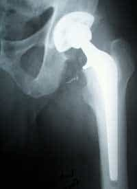
Cemented total hip prosthesis
Over the years, but not only, the joints lose elasticity and arthrosis phenomena can occur which cause pain, up to a poor mobility of the patient. In these cases, a hip prosthesis similar to the one visible in the radiograph to the side is used. The procedure itself is quite simple and takes about an… Continua a leggere Cemented total hip prosthesis
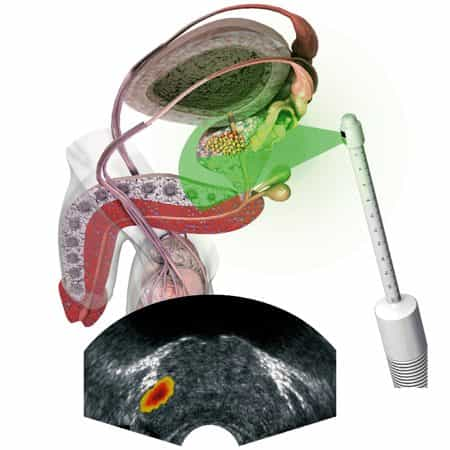
Early diagnosis of prostate cancer
Treating prostate cancer correctly is a race against time. By the time the patient feels the first symptoms, the disease has already spread. In this context, diagnostic techniques are of fundamental importance: CT, x-rays and magnetic resonance work well, but due to high costs they cannot become routine tests and sometimes they are not even… Continua a leggere Early diagnosis of prostate cancer
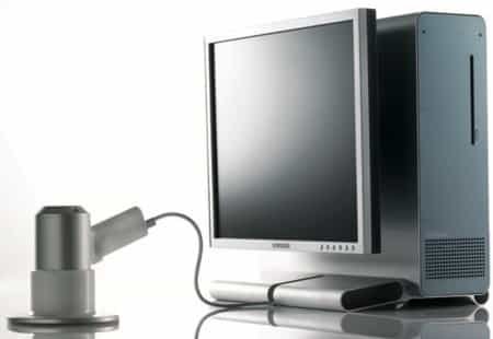
Imaging technique to diagnose melanoma
What you see above is the photo of SolarScan, the new generation of digital diagnostic devices to detect melanoma . Imaging techniques have literally revolutionized the dermatology sector, allowing specialist doctors quick access to data, a better study of the skin and above all the possibility of comparing reports made at different points in time.… Continua a leggere Imaging technique to diagnose melanoma